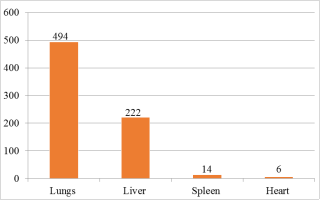
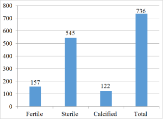
Hydatidosis is a parasitic zoonosis caused by the larval stage of Echinococcus granulosus, posing significant public health and economic concerns in camels. A cross-sectional study was conducted from November 2018 to April 2019 to determine the prevalence of camel hydatidosis and its associated risk factors in camels slaughtered at Akaki Municipal Abattoir, Addis Ababa, Ethiopia. A total of 364 camels were randomly selected and subjected to ante-mortem and post-mortem examination. Sex, age, body condition, and origin were recorded before thorough inspection of the lungs, liver, spleen, heart, and other organs for hydatid cysts. The overall prevalence of hydatidosis was 53.6% (195/364). Females (64.5%) and older camels (>10 years; 57.7%) showed significantly higher infection rates. Camels with medium body condition had a higher prevalence (59.4%) than those in good condition. Statistically significant associations (p = 0.00) were found between hydatidosis and sex, age, and body condition, while origin showed no significant association (p > 0.05). Among 239 infected organs, the lungs were the most frequently affected (60.7%), followed by the liver (34.4%), spleen (3.7%), and heart (1.2%). Of 736 collected cysts, 494 were found in the lungs and 222 in the liver, with 157 (21.3%) fertile, 545 (74.1%) sterile, and 122 (16.6%) calcified cysts. The high prevalence of hydatidosis highlights a lack of awareness in the camel-producing areas. Therefore, public education and targeted control strategies are recommended to reduce the burden of the disease.
| Published in | American Journal of Zoology (Volume 8, Issue 3) |
| DOI | 10.11648/j.ajz.20250803.11 |
| Page(s) | 60-65 |
| Creative Commons |
This is an Open Access article, distributed under the terms of the Creative Commons Attribution 4.0 International License (http://creativecommons.org/licenses/by/4.0/), which permits unrestricted use, distribution and reproduction in any medium or format, provided the original work is properly cited. |
| Copyright |
Copyright © The Author(s), 2025. Published by Science Publishing Group |
Hydatidosis, Camel, Prevalence, Akaki Abattoir, Ethiopia
Risk factors | Total camels examined | No of positive camels | Prevalence (%) | Chi-square value | P-Value |
|---|---|---|---|---|---|
Sex | |||||
Male | 212 | 97 | 45.8 | 12.47 | 0.00 |
Female | 152 | 98 | 64.5 | ||
Age | |||||
5-10 years | 47 | 12 | 25.5 | 17.05 | 0.00 |
>10 years | 317 | 183 | 57.7 | ||
Body condition score | |||||
Medium | 293 | 174 | 59.4 | 20.42 | 0.00 |
Good | 71 | 21 | 29.6 | ||
Origin | |||||
Borena | 181 | 96 | 53.00 | 2.17 | 0.338 |
Methara | 83 | 40 | 48.2 | ||
Minjar | 100 | 59 | 59 | ||
Total | 364 | 195 | 53.6 |
| [1] | FAOSTAT (Food and Agriculture Organization Statistics). (2011): Statistical database: |
| [2] | LDMPS. (Livestock Development Master Plan Study) (2006): Faculty of Veterinary Medicine Addis Ababa University. |
| [3] | Getahun, T. and Belay, K. (2002): Camel husbandry practices in Eastern Ethiopia: the case of Jigjiga and Shinile zone. Nomadic People. 6: 155-176. |
| [4] | Romazanvoc, F. (2001): Cestode zoonosis: Echnococcosis and Cysticercosis an emergent. And Global problem. IOS Press, Netherlands. Pp. 34-57. |
| [5] | Lahmar, S., Debbek, H., Zhang, P., McManus, A., Souissi, S., Chelly, M. and Torgerson, R. (2004): Transmission dynamics of the Echinoccus granulosus sheep-dog strain (GI genotype) in camel In Tunisia. Veterinary Parasitology, 121: 151-156. |
| [6] | Torgerson, P. and Budke, C. (2003): Echinococcosis-an international public health challenge. Research in Veterinary Science, 74(3): 191-202. |
| [7] | Seimenis, A. (2003): Overview of the epidemiological situation on echinococcosis in the Mediterranean region. Acta Trop, 85: 191-95. |
| [8] | Lightowlers, W., Flisser, A., Heath, D., Jensen, O. and Rolfe, R. (2000): Vaccination against Cysticercosis and Hydatid disease. Parasitology, 16: 191-196. |
| [9] | Wulamu, M, Hiroshi, Y, Yushito, S, Kazuhiro N, Minoru N, Lightowlers, M, Akira, I. (2002): usefulness of Hydatid cyst fluid of Echinococcus granulosus developed in mice with secondary infection for Serodiagnosis of Cystic Echinococcosis in humans. Clinical and Diagnostic Laboratory Immunology, 12: 573-576. |
| [10] | Macpherson, C., Bartholomot, B. and Frider, B. (1985): Application of ultrasound in diagnosis, treatment, epidemiology, public health and control of Echinoccus granulosus and E. multilocularis. Parasitology, 127(1): 21-35. |
| [11] | Kebede, N., Tilahun, G. and Hailu, A. (2009): Current status of bovine cysticercosis of slaughtered cattle in of slaughtered cattle in Addis Ababa Abattoir, Ethiopia. Tropical Animal Health and Production, 41: 290-294. |
| [12] | Thrusfield, M. (2007): Veterinary Epidemiology, 3rd edition UK. Blackwell science Ltd. Pp: 182-198. |
| [13] | Bulto, G., Yacob, H., Getachew, and T. and Hagos, A. (2013): Study on prevalence of Hydatidosis and cyst characterization in camels (camelus dromedaries) slaughtered at Akaki abattoir, Ethiopia. Journal of Veterinary Medicine and Animal Health, 5: 329-333. |
| [14] | Khan, B., Iqbal A., Riaz, M. (2003): Production and management of camels. University of Agriculture, Faisalabad, Pakistan. Pp. 152-156. |
| [15] | Faye, B., Bengoumi, M., Cleradin, A., Tabarani, A. and Chilliard, Y. (2001): Body condition score in dromedary camel: A too for management of reproduction. Emir. Journal of. Agricultural. Science. 13: 1-6. |
| [16] | Oostburg, B. F., Vrede, M. A. and Bergen, A. E. (2000): The occurrence of polycystic echinococcosis in Suriname. Annals of Tropical Medicine and Parasitology. 94, 247-252. |
| [17] | Adiselam, M. H., Mersha, Ch. K and Ismail W. J. (2014): Prevalence, Economic and Public Health Significance of Camel Hydatidosis in Dire Dawa Municipal Abattoir, Eastern Ethiopia. Acta Parasitologica Globalis, 5(2): 98-106. |
| [18] | Muskin, S., Moti, Y and Hailu, D. (2011): Infection rate, cyst fertility and larval viability of hydatid disease in camels (camelus dromedaries) from Borena, Kereyu and Harar areas of Ethiopia, Global Veternaria, 7: 518-522. |
| [19] | Ahmed, T. (1998): Preliminary investigation on major disease of camels of Eastern Ethiopia. Abattoir and field survey. DVM Thesis, Addis Ababa University, Faculty of Veterinary medicine, Debrezeit, Ethiopia. |
| [20] | Woldemeskel, M., Issa, A., Mersic, A and Potgieter, L. (2011): Investigation of parasitic disease of one- humped camel (Camelus dromedarius) in eastern Ethiopia. J. Camel Pract. Res, 8: 77-81. |
| [21] | Woubet, M (1987): A preliminary study of echinococcosis/hydatidosis in Hararghe region and the efficacy of Glinhslotoidus seeds against Echinoccus granulosus in pups infected experimentally with hydatid material. DVM thesis, Addis Ababa University, Faculty of VeterinaryMedicine, Debrezeit, Ethiopia. |
| [22] | Pandev, S., Ouhell, H and Ouchou, H. (1986): Hydatidosis in sheep, goat and Camelus dromedaries in Morocco. Annals of Tropical Medicine and Parasitology, 80: 525-529. |
| [23] | Njoroge, E. M., Mbithi, J. M., Gathuma, T. M., Wachira, S. R. and Magambo, J. K. (2000): Application of ultrasonography in prevalence studies of hydatid cysts in goats in north-western Turkana, Kenya and Toposaland, southern Sudan. Ond. Journal of Veterinary Research, 67: 251-255. |
| [24] | Mulatu, M., Biruk, M., Tasew, UH., Ashwani, K. (2013): hydatidosis in Eastern part of Ethiopia. Momona Ethiop. J. Sci. 5(1): 107-114. |
| [25] | Rokni, B. (2009): Echinococcosis /Hydatidosis in Iran. Iranian Journal of Parasitology, 4: 1-16. |
| [26] | Ahmadi, A. (2005): Hydatidosis in camels (Camelus dromedarius) and their potential role in the epidemiology of E. granulosus in Iran. Journal of Helminthology, 79: 119-125. |
| [27] | Abdul-salam, J. M., Farah, M. A. (1988): Hydatidosis in camels in Kuwait. Parasitology Research, 74: 267-270. |
| [28] | Bitsat, K. (2009): The prevalence of hydatidosis in jijiga municipal abattoir, DVM Thesis. Jimma University, Ethiopia. |
| [29] | Esatgil, M. and Tuzer, E. (2007): Prevalence of hydatidosis in slaughtered animals in Thrace, Turkey. Turkiye Parasitology Dergisi, 31: 43-45. |
| [30] | Himonas, C., (1987): The Fertility of Hydatid cyst in Food Animals in Greece. Helmenth, Zoonosis, Martin, Nijohoft, Publisher, Neitherland, pp: 12-18. |
APA Style
Tilahun, A., Kebede, A. (2025). Prevalence, Organ Distribution and Risk Factors Associated with Hydatidosis in Camels Slaughtered at Akaki Municipal Abattoir, Addis Ababa, Ethiopia. American Journal of Zoology, 8(3), 60-65. https://doi.org/10.11648/j.ajz.20250803.11
ACS Style
Tilahun, A.; Kebede, A. Prevalence, Organ Distribution and Risk Factors Associated with Hydatidosis in Camels Slaughtered at Akaki Municipal Abattoir, Addis Ababa, Ethiopia. Am. J. Zool. 2025, 8(3), 60-65. doi: 10.11648/j.ajz.20250803.11
@article{10.11648/j.ajz.20250803.11,
  author = {Ayansa Tilahun and Abriham Kebede},
  title = {Prevalence, Organ Distribution and Risk Factors Associated with Hydatidosis in Camels Slaughtered at Akaki Municipal Abattoir, Addis Ababa, Ethiopia
},
  journal = {American Journal of Zoology},
  volume = {8},
  number = {3},
  pages = {60-65},
  doi = {10.11648/j.ajz.20250803.11},
  url = {https://doi.org/10.11648/j.ajz.20250803.11},
  eprint = {https://article.sciencepublishinggroup.com/pdf/10.11648.j.ajz.20250803.11},
  abstract = {Hydatidosis is a parasitic zoonosis caused by the larval stage of Echinococcus granulosus, posing significant public health and economic concerns in camels. A cross-sectional study was conducted from November 2018 to April 2019 to determine the prevalence of camel hydatidosis and its associated risk factors in camels slaughtered at Akaki Municipal Abattoir, Addis Ababa, Ethiopia. A total of 364 camels were randomly selected and subjected to ante-mortem and post-mortem examination. Sex, age, body condition, and origin were recorded before thorough inspection of the lungs, liver, spleen, heart, and other organs for hydatid cysts. The overall prevalence of hydatidosis was 53.6% (195/364). Females (64.5%) and older camels (>10 years; 57.7%) showed significantly higher infection rates. Camels with medium body condition had a higher prevalence (59.4%) than those in good condition. Statistically significant associations (p = 0.00) were found between hydatidosis and sex, age, and body condition, while origin showed no significant association (p > 0.05). Among 239 infected organs, the lungs were the most frequently affected (60.7%), followed by the liver (34.4%), spleen (3.7%), and heart (1.2%). Of 736 collected cysts, 494 were found in the lungs and 222 in the liver, with 157 (21.3%) fertile, 545 (74.1%) sterile, and 122 (16.6%) calcified cysts. The high prevalence of hydatidosis highlights a lack of awareness in the camel-producing areas. Therefore, public education and targeted control strategies are recommended to reduce the burden of the disease.
},
 year = {2025}
}
TY  - JOUR
T1  - Prevalence, Organ Distribution and Risk Factors Associated with Hydatidosis in Camels Slaughtered at Akaki Municipal Abattoir, Addis Ababa, Ethiopia

AU  - Ayansa Tilahun
AU  - Abriham Kebede
Y1  - 2025/09/09
PY  - 2025
N1  - https://doi.org/10.11648/j.ajz.20250803.11
DO  - 10.11648/j.ajz.20250803.11
T2  - American Journal of Zoology
JF  - American Journal of Zoology
JO  - American Journal of Zoology
SP  - 60
EP  - 65
PB  - Science Publishing Group
SN  - 2994-7413
UR  - https://doi.org/10.11648/j.ajz.20250803.11
AB  - Hydatidosis is a parasitic zoonosis caused by the larval stage of Echinococcus granulosus, posing significant public health and economic concerns in camels. A cross-sectional study was conducted from November 2018 to April 2019 to determine the prevalence of camel hydatidosis and its associated risk factors in camels slaughtered at Akaki Municipal Abattoir, Addis Ababa, Ethiopia. A total of 364 camels were randomly selected and subjected to ante-mortem and post-mortem examination. Sex, age, body condition, and origin were recorded before thorough inspection of the lungs, liver, spleen, heart, and other organs for hydatid cysts. The overall prevalence of hydatidosis was 53.6% (195/364). Females (64.5%) and older camels (>10 years; 57.7%) showed significantly higher infection rates. Camels with medium body condition had a higher prevalence (59.4%) than those in good condition. Statistically significant associations (p = 0.00) were found between hydatidosis and sex, age, and body condition, while origin showed no significant association (p > 0.05). Among 239 infected organs, the lungs were the most frequently affected (60.7%), followed by the liver (34.4%), spleen (3.7%), and heart (1.2%). Of 736 collected cysts, 494 were found in the lungs and 222 in the liver, with 157 (21.3%) fertile, 545 (74.1%) sterile, and 122 (16.6%) calcified cysts. The high prevalence of hydatidosis highlights a lack of awareness in the camel-producing areas. Therefore, public education and targeted control strategies are recommended to reduce the burden of the disease.

VL  - 8
IS  - 3
ER  -
Department of Veterinary Medicine, School of Veterinary Medicine, Wollo University, Dessie, Ethiopia;Department of Veterinary Medicine, School of Veterinary Medicine, Wollega University, Nekemte, Ethiopia
Department of Veterinary Medicine, School of Veterinary Medicine, Wollo University, Dessie, Ethiopia;Department of Veterinary Medicine, School of Veterinary Medicine, Wollega University, Nekemte, Ethiopia